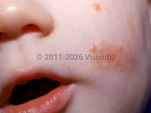
Benign cephalic histiocytosis (BCH) is a rare non-Langerhans histiocytosis that typically occurs between 2 and 34 months of age and, less commonly, up to the age of 5 years. Approximately 50% of cases begin between the ages of 5 and 12 months. It affects males and females equally. The condition is characterized by asymptomatic, raised, reddish-yellow papules, 2-4 mm in diameter. Papules initially develop on the head in all cases, most often the cheeks, eyelids, forehead, and ears. Lesions may extend to involve the neck and upper trunk. Affected children will always have multiple lesions. Unlike other forms of histiocytoses, mucosa and viscera are not involved.
Involution of lesions occurs spontaneously over the ages of 2-8 years, often with residual postinflammatory hyperpigmentation that persists indefinitely. There have been no reported associated systemic diseases or reports of visceral involvement. Some experts consider BCH to be a childhood variant of generalized eruptive histiocytoma, and some reports suggest that BCH may have overlapping clinical and histologic features with juvenile xanthogranuloma (JXG). There have been reports of BCH transforming later into JXG, further establishing this theoretical overlap. BCH may be difficult to differentiate clinically from JXG, but key differentiating features are that BCH lesions tend to be flatter and are mainly on the head and neck.
Benign cephalic histiocytosis in Child
Alerts and Notices
Important News & Links
Synopsis

Codes
ICD10CM:
D76.3 – Other histiocytosis syndromes
SNOMEDCT:
255192005 – Benign cephalic histiocytosis
D76.3 – Other histiocytosis syndromes
SNOMEDCT:
255192005 – Benign cephalic histiocytosis
Look For
Subscription Required
Diagnostic Pearls
Subscription Required
Differential Diagnosis & Pitfalls

To perform a comparison, select diagnoses from the classic differential
Subscription Required
Best Tests
Subscription Required
Management Pearls
Subscription Required
Therapy
Subscription Required
References
Subscription Required
Last Updated:11/18/2019
Benign cephalic histiocytosis in Child